United States Military Tests Wavefront-Guided PRK

With more than 30,000 to 40,000 members of the United States military choosing laser vision correction procedures to improve their vision each year, there is an emphasis on clinical research to examine the efficacy and safety of various procedures available.

A somewhat novel laser vision correction technique, called wavefront-guided PRK (Photorefractive Keratectomy), was recently studied by the United States Navy.

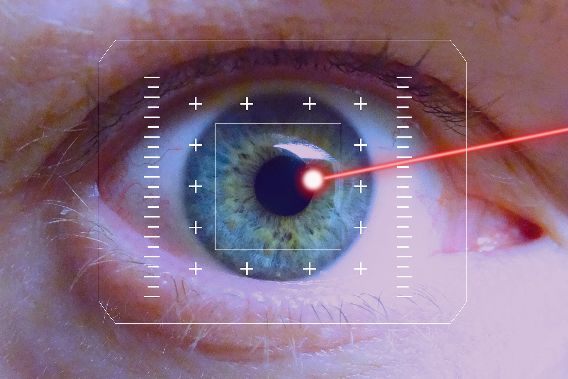
Wavefront-guided PRK implements the newest computer-generated mapping technology to generate a digital image of the patient’s eye. Once the eye is mapped out, the laser applied to the cornea’s surface is guided by the three-dimensional model to improve the eye’s focus based on the unique visual characteristics of the patient’s eye. Even the tiniest structure issues affecting eyesight are revealed through Wavefront-Guided PRK.

The development of wavefront vision mapping allows visual disorders to be measured, which otherwise could not be identified in the past. Laser diagnostic systems are setting a new standard by providing an exact level of measurement and correction. This feat was impossible with earlier systems.

Led by the U.S. Naval Medical Center, 167 patients enrolled had vision prescriptions of up to -8D of nearsightedness and up to -4D of astigmatism. The patients underwent wavefront-guided PRK. Research findings showed:

  • Patient satisfaction was very high, with 98.8 percent reporting entirely or very satisfied with their vision
  • 99.4 percent of patients tested at 20/20 or better vision six months following the procedures
  • 91.9 percent of patients tested 20/16 vision six months after the procedure
  • Fewer than four percent of patients reported night vision symptoms six months post-op
  • Less than one percent experienced adverse reactions

This novel research shows a massive body of evidence supporting laser vision correction procedures proving safety and effectiveness. 

Rohr Eye & Laser Center offers the most advanced technology available to provide personalized and extraordinary care to our patients. Whether your goal is to maintain or improve your natural vision, we are here to help you. Call us at 877-579-0202 or visit https://www.michiganlasik.com/ to schedule an appointment today.

Written by the digital marketing staff at Creative Programs & Systems: www.cpsmi.com.

Retinal Transplants Ready for Human Trial

A major cause of hereditary human blindness, Retinitis Pigmentosa (RP), has been resolved in rat trials by researchers at the RIKEN Center for Biosystems Dynamics Research (BDR) in Japan. Degenerated rat retinas received transplants and removal of grafted cells, which resulted in improved connections to host retinas and better light-responsiveness.

Study leader Michiko Mandai said, “We have previously reported work using mouse embryonic- and induced pluripotent stem cell – (iPES)-derived retinas in a mouse model of retinal degeneration.”

However, the deputy project leader in the Laboratory for Retinal Regeneration at RIKEN BDR said, “As far as we know, this is the first report of the timed removal of particular grafted cells being shown to improve retinal function in an animal model using tissue from human stem cells.”

Since the retinal sheets were created from human stem cells, the novel feat can now be tested in human clinical trials, according to the study authors in the January 21, 2022 edition of iScience.

“Since retinal ganglion cells form optic nerves to send the signal to the brain, a higher light responsiveness could theoretically lead to improved sensitivity to light or better resolution in visual function. Genetic modification in human stem cell-derived retinas showed a substantial functional improvement [in light responsiveness], compared with wild-type graft retinas. Depending on the retinal network of the transplanted area, light responsiveness of retinal ganglion cells may be correlated with increased signaling to the brain, so could lead to an increased light sensitivity or better resolution. Additionally, we were able to observe detailed host-graft synapse formation in the absence of graft bipolar cells, which previously was difficult. A frequently asked question in retinal cell therapies concerns whether retinal ganglion cell responses are really originated from the graft photoreceptors. The presence of multiple synaptic markers at the host-graft cell contact site suggests formation of some synapses between these cells and indicates that graft photoreceptor light responses may well be transmitted to host retinal cells. We can now move forward to applying this strategy in clinical studies, which we expect to improve clinical outcomes and be generally useful in stem cell-based therapies targeting retinal degeneration. If the clinical grade ISL1-deleted stem cell-derived retinas can first pass extensive safety testing, including for tumorgenicity, we estimate that this may take 4-5 years. While we are currently targeting end-stage disease eyes with stem cell based therapy, we think this strategy could also be applied to those with remaining central vision, to help increase the pericentral visual field or delay disease progression,” Mandai said.

Rohr Eye & Laser Center offers the most advanced technology available to provide personalized and extraordinary care to our patients. Whether your goal is to maintain or improve your natural vision, we are here to help you. Call us at 877-579-0202 or visit https://www.michiganlasik.com/ to schedule an appointment today.

Written by the digital marketing staff at Creative Programs & Systems: www.cpsmi.com.

Dried Goji Berries Can Prevent Age-Related Vision Loss

A small, randomized trial by the University of California, Davis, found that people who regularly eat a small serving of dried goji berries might see prevention or delay of age-related macular degeneration (AMD) in healthy, middle-aged people.

Affecting over 170 people globally and roughly 11 million throughout the United States, AMD is the leading cause of vision loss in elderly people.

Glenn Yiu, a co-author of the study and associate professor in the Department of Ophthalmology and Vision Sciences, said, “AMD affects your central field of vision and can affect your ability to read or recognize faces.”

Research concluded that 13 study participants ages 45 to 65 increased the density of protective eye pigments after eating 28 grams (a handful) of goji berries five times per week for 90 days. Eating goji berries increased eye pigments lutein and zeaxanthin, which filter out harmful blue light, provide support through antioxidant protection, and protect aging eyes.

Lead author Xiang Li, a doctoral candidate in the Nutritional Biology Program, said, “Lutein and zeaxanthin are like sunscreen for your eyes. The higher the lutein and zeaxanthin in your retina, the more protection you have. Our study found that even in normal healthy eyes, these optical pigments can be increased with a small daily serving of goji berries.”

Goji berries are commonly used in Chinese medicine to “brighten” eyes through lutein and zeaxanthin. Dietary supplements containing Vitamin C, Vitamin E, zinc, copper, lutein, and zeaxanthin are used to treat intermediate stages of AMD.

“Our study shows goji berries, which are a natural food source, can improve macular pigments of healthy participants beyond taking high-dose nutritional supplements,” said Yiu. “The next step for our research will be to examine goji berries in patients with early-stage AMD.”

Rohr Eye & Laser Center offers the most advanced technology available to provide personalized and extraordinary care to our patients. Whether your goal is to maintain or improve your natural vision, we are here to help you. Call us at 877-579-0202 or visit https://www.michiganlasik.com/ to schedule an appointment today.

Written by the digital marketing staff at Creative Programs & Systems: www.cpsmi.com.

Declining Eyesight Improved with Red Light Therapy

A novel study by researchers at UCL found that three minutes of exposure to deep red light once a week can vastly improve deteriorating eyesight.

The study, published in Scientific Reports, outlines how using longwave deep red light in the morning activated mitochondria cells in the human retina, repairing declining vision. Previous studies focused on higher energy levels and different times of the day to perform the therapy.

Overall, researchers discovered an average of 17 percent improvement in color contrast vision when exposed to three minutes of deep red light in the morning, which lasted at least a week. The same test, performed in the afternoon, showed no vision improvement.

Researchers’ findings exhibit a breakthrough for eye health which could lead to more affordable home-based therapies. Millions of people suffer worldwide from naturally declining vision, which could be improved with just three minutes of red light therapy every morning.

Professor Glen Jeffery, UCL Institute of Ophthalmology, lead author, said, “We demonstrate that one single exposure to longwave deep red light in the morning can significantly improve declining vision, which is a major health and wellbeing issue, affecting millions of people globally. This simple intervention applied at the population level would significantly impact on quality of life as people age and would likely result in reduced social costs that arise from problems associated with reduced vision.”

Working with Planet Lighting UK, Jeffery aims to produce 670nm infrared eyewear at an affordable price; other LED devices made specifically for the same purpose can cost more than $20,000.

Jeffery said, “The technology is simple and very safe; the energy delivered by 670nm long wave light is not that much greater than that found in natural environmental light. Given its simplicity, I am confident an easy-to-use device can be made available at an affordable cost to the general public. In the near future, a once a week three-minute exposure to deep red light could be done while making a coffee, or on the commute listening to a podcast, and such a simple addition could transform eye care and vision around the world.”

Rohr Eye & Laser Center offers the most advanced technology available to provide personalized and extraordinary care to our patients. Whether your goal is to maintain or improve your natural vision, we are here to help you. Call us at 877-579-0202 or visit https://www.michiganlasik.com/ to schedule an appointment today.

Written by the digital marketing staff at Creative Programs & Systems: www.cpsmi.com.

Blurry Vision after Eating

If you have ever stood up after eating and realized your vision is slightly blurry, you might have experienced a brief blood sugar level spike. If this happens to you more often than not, you may want to schedule a visit with your eye care professional to see whether you have an underlying condition (such as diabetes), which causes these spikes.

Excessively high blood sugar levels generally cause people to be diagnosed with diabetes. When food is digested more rapidly, blood sugar levels can spike. Fluid can build up in the eyes, resulting in blurry vision.

In the eye, the cornea and natural crystalline lens work together to focus light into the retina, resulting in clear vision. To accommodate focusing on objects near or far, the lens changes its shape. In some instances, high blood sugar can cause the eye to swell and cause the focus to be off.

Some foods high in sugar and likely to spike blood sugar include white rice, pasta, cereals, potatoes, pop, fruit juices, candy, and baked goods.

Blurry vision doesn’t always signify diabetes. Other conditions include:

  • Carotid stenosis
  • Migraines
  • Medications
  • Eyestrain
  • Pink eye (conjunctivitis)
  • Keratitis
  • Glaucoma
  • Macular degeneration

Symptoms other than blurry vision typically accompany these conditions.

Rohr Eye & Laser Center offers the most advanced technology available to provide personalized and extraordinary care to our patients. Whether your goal is to maintain or improve your natural vision, we are here to help you. Call us at 877-579-0202 or visit https://www.michiganlasik.com/ to schedule an appointment today.

Written by the digital marketing staff at Creative Programs & Systems: www.cpsmi.com.

New Changes in FSA Make LASIK More Affordable

A Flexible Spending Account (FSA) is tax-exempt money you can set aside from your paycheck to pay out-of-pocket expenses. Throughout the year, the level you contribute is consistent and even. The maximum amount you can contribute in 2021 is $2,750.

If you have been considering getting LASIK, your FSA could be used to cover the cost. Recent legislation has made it possible for employers to extend the deadline for all 2021 FSA spending. Thanks to this new extension, employees can use their FSA to pay for LASIK.

There are a few limitations when it comes to FSAs, including:

  • Pretax funds can only be spent on qualified health and medical expenses
  • The current limit that can be used in an FSA account is $2,750
  • Any remaining cash in the FSA after December 31 is forfeited – all funds must be used within the calendar year
  • Grace periods and the ability to carry over a portion of the funds do exist

FSA was limited due to the global coronavirus pandemic, making spending a challenge. The 2021 FSA deadlines were extended to compensate for this pause in healthcare. Now, the entire amount of FSA can be carried through December 31, 2022.

To use your FSA to pay for LASIK, contact your employer’s HR department to determine whether they have opted to implement the flexible deadlines. Check your balance to calculate how much money you will have available. Schedule a consultation ASAP, and soon enough, you can sigh in relief…no more glasses or contacts after LASIK!

Thinking about LASIK? There’s no better time to achieve your dreams! Contact Rohr Eye & Laser Center today to get started.

Rohr Eye & Laser Center offers the most advanced technology available to provide personalized and extraordinary care to our patients. Whether your goal is to maintain or improve your natural vision, we are here to help you. Call us at 877-579-0202 or visit https://www.michiganlasik.com/ to schedule an appointment today.

Written by the digital marketing staff at Creative Programs & Systems: www.cpsmi.com.

Better Verbal Communication for Superior Glaucoma Treatment

Patient-physician discussions are vital for treatment and education, but a new study shows that better communication may also be a valuable tool for determining the severity of glaucoma disease.

“One specific challenge in communicating about glaucoma is that the treating physician doesn’t fully grasp what the patient sees and the words they might use to describe these visual symptoms,” the researchers said. Better communication helps physicians understand the patient’s vision concerns.

One hundred seventy adults diagnosed with glaucoma were analyzed. Questionnaires rated visual symptoms, and the most notable cases were identified. Eye damage was reported in patients who experienced blurry vision, sensitivity to light, glare, cloudy vision, and poor peripheral vision. Those who exhibited the most significant variance in vision (43 percent) were people who complained of little peripheral vision.

Researchers noted there was a 62 percent variance in eye damage. Symptoms described by patients coincided with glaucoma. The information can help judge glaucoma in clinical settings.

“These symptoms suggest specific terms that are likely to reflect glaucoma damage, which can be used when communicating with patients,” said researchers.

“Missing patches can point to glaucomatous scotomas, cloudy vision can be understood as the manifestation of contrast deficits which underlie glaucoma, and worsening vision may reflect eyesight severity because of a greater rate of worsening in severe disease. Reporting worsening vision may also reflect the clinically-observed tendency of subjective vision to fluctuate more in advanced glaucoma, resulting in days of worsening, though some corresponding days of improvement would also be present,” said researchers.

Good communication can help physicians understand patient symptoms, strengthen relationships between the two, and enable patients to understand the impact of eyesight diseases on their life.

The researchers said, “Understanding what symptoms patients with severe glaucoma experience is uniquely important, as they may feel frustrated completing visual testing or lose faith in treatment due to their limited vision. If physicians demonstrate an understanding of what specific symptoms are related to severe glaucoma and the impact on a patient’s vision, they may be able to better develop therapeutic alliances with patients.”

Rohr Eye & Laser Center offers the most advanced technology available to provide personalized and extraordinary care to our patients. Whether your goal is to maintain or improve your natural vision, we are here to help you. Call us at 877-579-0202 or visit https://www.michiganlasik.com/ to schedule an appointment today.

Written by the digital marketing staff at Creative Programs & Systems: www.cpsmi.com.

Blind Woman Sees Shapes Thanks to Scientists

A team of researchers from the University Miguel Hernandez (Spain), the Netherlands Institute of Neuroscience (Netherlands), and the John A. Moran Eye Center at the University of Utah (USA) recently published details about how they were able to successfully restore vision in a woman using a brain implant.

The scientists oversaw a series of tests with the blind volunteer to eventually recreate her vision. First, a neurosurgeon implanted a microelectrode array into her visual cortex, which was composed of 100 microneedles intended to stimulate neurons near the electrodes. The woman wore eyeglasses outfitted with a mini video camera which translated the data collected and sent to the brains electrodes. Finally, the brain simulated the image by surrounding neurons to produce white areas of light known as “phosphenes.”

A former science teacher, the woman had been blind for 16 years. No complications were seen post-op, and scientists concluded the implant did not affect brain function. 

Lines, shapes, and letters were visible to the blind woman. The researchers developed a game to help her strengthen and recognize the different phosphene shapes. Because of her extensive involvement and unique insight, the woman became a co-author of the study.

Professor Eduardo Fernandez said, “These results are very exciting because they demonstrate both safety and efficacy and could help to achieve a long-held dream of many scientists, which is the transfer information from the outside world directly to the visual cortex of blind individuals, thereby restoring a rudimentary form of sight. Although these preliminary results are very encouraging, we should be aware that there are still a number of important unanswered questions and that many problems have to be solved before a cortical visual prosthesis can be considered a viable clinical therapy.”

Professor R. A. Normann, a study co-author, said, “It could allow them to identify a person, doorways, or cars. It could increase independence and safety. That’s what we’re working toward.”

Professor P. Roelfsema, a co-author in the study, said, “This new study provides proof-of-principle and demonstrates that our previous findings in monkey experiments can be translated to humans. This work is likely to become a milestone for the development of new technologies that could transform the treatment of blindness.”

Rohr Eye & Laser Center offers the most advanced technology available to provide personalized and extraordinary care to our patients. Whether your goal is to maintain or improve your natural vision, we are here to help you. Call us at 877-579-0202 or visit https://www.michiganlasik.com/ to schedule an appointment today.

Written by the digital marketing staff at Creative Programs & Systems: www.cpsmi.com.

Research Proves LASIK Adverse Events are Rare

Ophthalmology is a scientific field that is continuously performing research and studies based on the human eye. LASIK eye surgery is an imperative aspect of ophthalmology that presents patients with more benefits than risks.

A recent study published in the American Journal of Ophthalmology outlined researchers’ data analysis from roughly 37,000 laser vision correction patients who underwent either LASIK (31,921) or PRK (5,016) between July 2014 and June 2016. Data analyzed included eye examinations prior to the surgery, procedure reports, and follow-up exams.

Data on adverse events were also recorded, such as those that negatively impacted patients’ visual outcomes. Out of 37,000 patients, 34 experienced adverse side effects; 70 percent were characterized as severe. With less than one percent of patients experiencing a significant unfavorable result, each procedure showed:

  • LASIK (31,921 patients): 0.4 percent of patients experienced unfavorable results
  • PRK (5,016 patients): 0.7 percent of patients experienced unfavorable results

Of those with serious adverse effects, roughly 80 percent still achieved 20/20 (or higher) vision without glasses or contacts.

With over 20 million procedures performed throughout the United States over 20 years, research papers number greater than 7,000. Experts study vision correction effects in-depth to ensure they are safe and effective for patients. Based on the large number of patients country-wide, LASIK and PRK are excellent, viable options to improve vision for suitable candidates. 

To improve your vision through LASIK, contact the ophthalmology experts at Rohr Eye & Laser Center today.

Rohr Eye & Laser Center offers the most advanced technology available to provide personalized and extraordinary care to our patients. Whether your goal is to maintain or improve your natural vision, we are here to help you. Call us at 877-579-0202 or visit https://www.michiganlasik.com/ to schedule an appointment today.

Written by the digital marketing staff at Creative Programs & Systems: www.cpsmi.com.

Five Most Common LASIK Questions

Laser vision correction is a viable option for many people struggling with dry eyes, foggy glasses when wearing masks, contact lens fatigue from too much screen time, and plain ol’ aging. These issues are a nuisance and can be easily remediated with LASIK.

Before most people make the jump, they ask whether LASIK is safe. The answer is simple: yes, LASIK is safe. Over 7,000 clinical studies have investigated – and verified – its safety and effectiveness. Continue reading to discover more questions and answers to the most common questions.

How does LASIK work? LASIK essentially sculpts a contact lens onto the eye’s surface, giving the patient vision as if they are looking through a contact lens without the uncomfortable feeling of a lens.

What does LASIK correct? LASIK has corrected vision conditions, including myopia (nearsightedness), hyperopia (farsightedness), and astigmatism.

Am I a good candidate? The best way to discover whether you are a good candidate for LASIK is to have a complete examination.

How many patients have 20/20 vision after LASIK? Many published studies show that roughly 96 to 99 percent of patients attain 20/20 vision or better after a laser procedure.

How long does LASIK last? Typically, the vision correction outlasts the patient. However, since eyes age with age, it might fluctuate a bit.

To learn more about how LASIK can improve your vision, contact the experts at Rohr today.

Rohr Eye & Laser Center offers the most advanced technology available to provide personalized and extraordinary care to our patients. Whether your goal is to maintain or improve your natural vision, we are here to help you. Call us at 877-579-0202 or visit https://www.michiganlasik.com/ to schedule an appointment today.

Written by the digital marketing staff at Creative Programs & Systems: www.cpsmi.com.